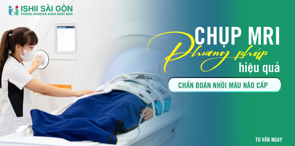
Cerebral infarction is one of the primary causes of stroke and mortality in many countries worldwide. This is particularly true in Vietnam, where the number of patients with acute cerebral infarction is rapidly increasing each year. Therefore, timely diagnosis and treatment are crucial factors in reducing the mortality rate due to acute cerebral infarction. In this article, we will explore the role of MRI in diagnosing acute cerebral infarction and the MRI imaging process at Ishii Saigon Clinic.
What is Acute Cerebral Infarction?

Acute cerebral infarction is a condition where a blood vessel in the brain is blocked or ruptured, leading to a reduction or complete loss of blood supply to a specific area of the brain. Consequently, cells in the affected area do not receive enough oxygen and nutrients, causing damage and cessation of function. As a result, brain cells may die, leading to a decrease in their functionality.
Acute cerebral infarction typically occurs due to a blood clot (thrombosis) or an embolus from another part of the body that blocks a small blood vessel in the brain. Additionally, it can also result from a gas bubble or a piece of debris flowing through a missed blood vessel.
Symptoms of acute cerebral infarction include severe headache, dizziness, loss of ability to move or speak, numbness, blurred vision, and confusion. These symptoms can vary depending on the location and extent of damage to the brain's blood vessels. If not detected and treated promptly, acute cerebral infarction can pose life-threatening risks and lead to severe complications.
Advantages of MRI in the diagnosis of acute cerebral infarction
MRI (Magnetic Resonance Imaging) is a medical imaging technique that uses radio waves and magnetic fields to create detailed images of the body. Specifically, MRI is highly beneficial in diagnosing acute cerebral infarction due to its precise scanning capability and non-invasive nature, without impacting the patient's body.

One of the key advantages of MRI is its ability to allow physicians to visualize structures and tissues within the brain with higher resolution compared to other imaging methods. This enables doctors to clearly and accurately identify blocked or ruptured blood vessels, brain injuries, and other components within the brain.
Additionally, MRI does not use X-rays and does not cause any adverse effects on the body, helping to minimize risks for patients. This is particularly important for patients at risk of allergies or those who cannot tolerate X-rays.
MRI Procedure at Ishii Sai Gon Medical Clinic
Ishii Sai Gon Clinic is known as one of the leading medical centers in Vietnam for utilizing the 3.0 Tesla MRI machine to diagnose and treat neurological disorders. The MRI examination at Ishii Sai Gon typically takes about 15 minutes, providing accurate diagnostic results that enable doctors to propose appropriate treatment plans for patients.
Procedure for MRI at Ishii Saigon Clinic includes the following steps:
Step 1: Preparation
Before starting the MRI procedure, patients are asked to change into medical attire and lie still on the MRI bed. Patients should avoid bringing fragile items, watches, electronic accessories, or metal objects into the MRI room. If any are present, they will be asked to remove them before entering the imaging room.
Step 2: Endoscopy
In some cases, in addition to conventional MRI scans, doctors may also request an endoscopy to obtain clearer images of the affected area. This helps doctors examine and accurately assess the patient's condition.
Step 3: MRI Scan
After preparation, the doctor will take the patient into the MRI room. Here, the patient will be asked to lie still for approximately 30-45 minutes, depending on the purpose of the MRI scan. Throughout the scan, the patient will remain in the same position on the bed and move into the MRI machine.
Step 4: Evaluation and Diagnosis
After completing the scanning process, the doctors will review and evaluate the MRI images to make an accurate diagnosis. The results will be communicated to the patient as soon as possible for timely treatment.
Considerations when performing MRI for diagnosing cerebral infarction
Similar to other MRI machines, the 3.0 Tesla machine also has specific requirements that need to be followed during the scanning process. Therefore, patients should take note of the following points after deciding to undergo an MRI to ensure a safe and effective procedure:
- Avoid wearing electronic accessories into the MRI room, as mentioned above.
- Remove all metal items (such as gold, watches, necklaces, artificial nails, etc.) before entering the MRI room.
- Avoid bringing fragile items.
- Do not use cosmetics, skincare products, or perfume before entering the room.
- If using anticoagulant medications (such as aspirin), inform the doctor before undergoing the MRI procedure.
MRI 3.0 Tesla imaging at Ishii Saigon Clinic enhances the diagnosis of cerebral infarction

The MRI 3.0 Tesla machine is one of the most advanced technologies used for diagnosing and treating brain-related diseases in Vietnam today. Particularly, Ishii Saigon Clinic has successfully utilized this machine for the accurate diagnosis and treatment of acute cerebral infarction, ensuring high precision without harm to patients.
This is crucial in reducing the mortality rate from acute cerebral infarction and also improves the quality of life for those affected by this condition. MRI imaging using the 3.0 Tesla machine at Ishii Saigon Clinic not only serves as a diagnostic method but also aids doctors in monitoring and evaluating the patient's condition throughout the treatment process.
Conclusion
The above information highlights the role of MRI in diagnosing acute cerebral infarction, the procedure for performing MRI at Ishii Saigon Clinic, and considerations during this process. It is evident that employing modern technology such as the 3.0 Tesla MRI machine not only benefits doctors in diagnosis and treatment but also enhances the quality of life for individuals suffering from acute cerebral infarction. Therefore, it is important to recognize and prioritize the role of MRI in diagnosing and treating neurological conditions to safeguard the health of oneself and those around us.


