


Magnetic Resonance Imaging (MRI) is an advanced imaging diagnostic technique that uses magnetic fields and radio waves to create detailed images of the brain and other parts of the body. MRI does not use X-rays, making it safer for patients compared to other imaging methods such as computed tomography (CT) scans.
What is Magnetic Resonance Imaging (MRI)?
MRI works based on the principle of nuclear magnetic resonance of hydrogen atoms in the body. When the body is placed in a strong magnetic field, hydrogen nuclei align in the same direction. Radio waves are then used to stimulate these hydrogen nuclei, causing them to deviate from their original positions. As the hydrogen nuclei return to their original positions, they emit signals. These signals are detected and processed by a computer to create detailed images of the body's internal structures.
Advantages of MRI:
- Provides more detailed and clearer images compared to other imaging diagnostic methods.
- Does not use X-rays, therefore safer for patients, especially children and pregnant women.
- Can diagnose various conditions, including brain tumors, spinal cord tumors, strokes, and musculoskeletal disorders.
Disadvantages of MRI:
- Higher cost compared to other imaging diagnostic methods.
- The MRI scanning process may take longer compared to CT scans.
- Some patients may feel uncomfortable due to the loud noise while lying inside the MRI machine.
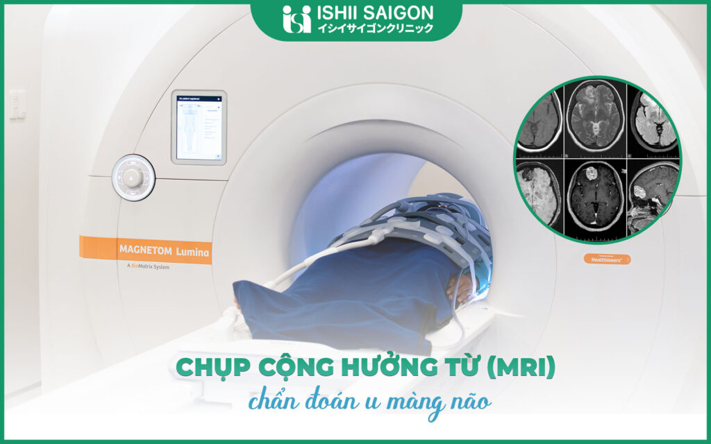
How to diagnose a brain tumor using magnetic resonance imaging (MRI)
MRI is the most effective imaging diagnostic method for detecting brain tumors. MRI can help determine the location, size, and shape of the tumor mass, as well as its relationship to surrounding tissues.
The process of diagnosing a brain tumor with MRI:
- The patient is placed into the MRI machine and lies still throughout the scanning process.
- The technician will use radio waves to generate images of the brain.
- The MRI scanning process can take from 30 minutes to 1 hour.
The doctor will rely on the MRI images to diagnose the brain tumor and determine the appropriate treatment method.
Magnetic Resonance Imaging and consultation with a Japanese doctor at Ishii Saigon.
At Ishii Saigon Japanese Clinic, patients can undergo MRI scans and receive consultations from experienced Japanese specialists. The Japanese doctors at Ishii Saigon are well-trained in brain tumor imaging diagnosis and can provide patients with precise and appropriate medical advice.
Advantages of undergoing MRI and receiving consultations at Ishii Saigon:
- State-of-the-art equipment and machinery imported from Japan.
- Experienced and dedicated specialist doctors.
- Attentive and professional customer care services.
The Ishii Saigon Japanese Clinic is a reputable and high-quality healthcare facility specialized in diagnosing and treating neurological disorders, including brain tumors. Equipped with state-of-the-art machinery and equipment imported from Japan, the clinic boasts a team of experienced and dedicated specialist doctors who are committed to providing excellent patient care.

To receive consultation and schedule an MRI scan for diagnosing brain tumors, please contact:
Ishii Sai Gon - Japanese Medical Clinic
Address: 616A Nguyễn Chí Thanh, Ward 4, District 11, Ho Chi Minh City (Opposite the main gate of Cho Ray Hospital).
Phone number 1900 636 079 – 0977 564 616
Website: www.ishiisaigon.vn